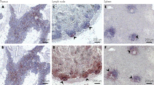
Figure 2 Peripheral lymphoid organ immunohistochemistry. (A,B) The thymus from SD600 has fewer CD4+ (A, anti‐OPD4) than CD8+ cells (B, anti‐CD8). (C,D) A hilar lymph node from SD600 showing depletion of follicular B (C, arrows, anti‐CD20) and T cells and aberrant T cell localisation (D, arrows, anti‐CD3). (E,F) Decreased number of B (E, arrow, anti‐CD20) and T cells (F, arrow, anti‐CD3) in splenic tissue from SD600.

An official website of the United States government
Here's how you know
Official websites use .gov
A
.gov website belongs to an official
government organization in the United States.
Secure .gov websites use HTTPS
A lock (
) or https:// means you've safely
connected to the .gov website. Share sensitive
information only on official, secure websites.
