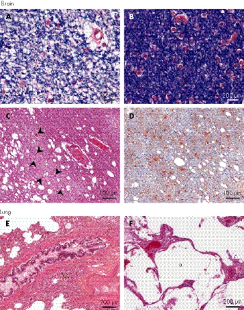
Figure 4 Histopathology and immunohistochemistry of the brain and lung. Brain: Focal paucity of myelin in the cerebral white matter from SD600, as shown by reduced (A) and normal (B) luxol fast blue staining. (C) Lacunar infarction with swollen axons (arrowheads) in the white matter (SD840, haematoxylin and eosin (H&E) stain). (D) Gliosis in the infarcted white matter (SD840, anti‐glial fibrillary acidic protein). Lungs: (E) Bronchial smooth muscle hyperplasia (SD600, H&E stain). (F) Dilated airspaces (a) in the lung (SD840, H&E stain).

An official website of the United States government
Here's how you know
Official websites use .gov
A
.gov website belongs to an official
government organization in the United States.
Secure .gov websites use HTTPS
A lock (
) or https:// means you've safely
connected to the .gov website. Share sensitive
information only on official, secure websites.
