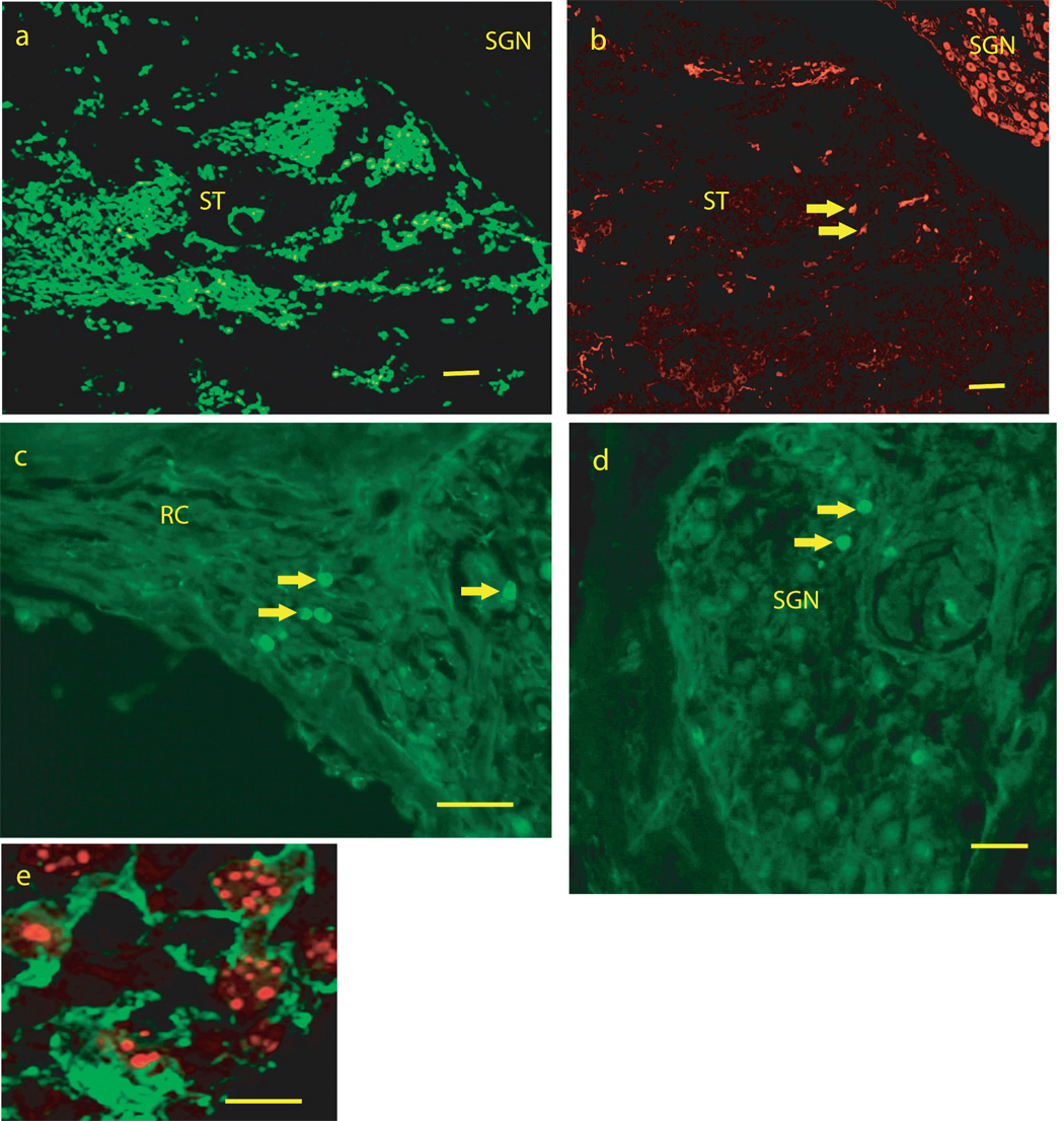
FIGURE 2.
Photomicrographs from mid-modiolar cryostat sections through the guinea pig cochlea at two weeks following placement of mouse embryonic stem cells into the scala tympani/modiolus. 2a: low magnification image from scala tympani in the basal turn of the cochlea spiral showing enhanced green fluorescent protein (eGFP) fluorescence. 2B: An adjacent section to the section in 2a immunostained for TUJ1, showing TUJ1 immunostaining of spiral ganglion neurons (SGN) in Rosenthals canal and of cells (see arrows for examples) in scala tympani (ST). Note that there is little or no overlap of eGFP fluorescence in 1a and TUJ1 immunofluoresence in 1b. Figures 2c and d show eGFP fluorescent cells (arrows for examples) among SGN (2c) and SGN peripheral processes (2d) in Rosenthals canal. 1e shows co-labeling for TUJ1 immunostaining (green and cytoplasmic) and in situ hybridization labeling of mouse genomic DNA (red, punctuate and nuclear) in cells in scala tympani of the basal turn of the cochlear spiral. SGN = region of Spiral Ganglion Neurons, RC = region of SGN peripheral processes in Rosenthals Canal, ST = Scala Tympani. Bars for 1 a–c = 100 microns, 1d = 50 microns, 1 e = 10 microns.

