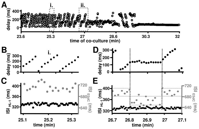
Figure 2. Delay of AP propagation between HL-1 cells and ESdC.
(A) For every excitation cycle of the HL-1 monolayer the delay between HL-1 and ESdC activity was determined during a period of independent activity (i.) and a period of transient synchronization (ii.). In i. the delay between HL-1 and ESdC activity is non-stationary (B). Separate analysis of the ESdCs and HL-1 spikes ISI revealed beating frequencies comparable to ESdCs and HL-1 cells in homocellular culture respectively (C). During transient synchronization (ii), the delay between the HL-1 cells and the ESdCs activity transiently stabilizes (D). At this point of time the ESdC’s ISI is an integral value of the HL-1’s ISI (E).

