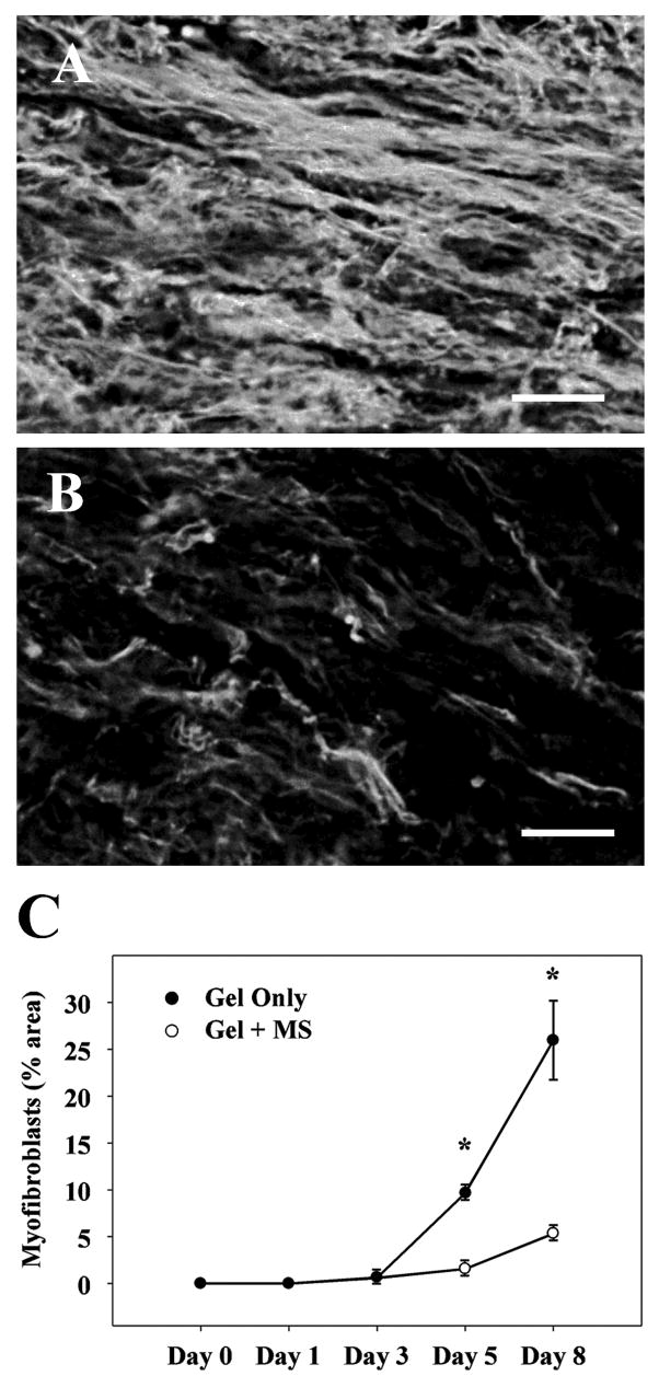
Fig. 3. Myofibroblast accumulation in the healing cutaneous wound.
Wounds received twice-daily treatment with IntraSite Gel (Smith+Nephew, Hull, United Kingdom) alone or gel infused with 5 mM morphine sulfate (MS). (A, B) αSMA-ir cells in granulation tissue of healing wounds 8 days following wounding. Fluorescence photomicrographs of gel-only (A) and morphine-treated (B) wounds. Note a significant decrease in myofibroblast density in morphine-treated wounds as compared to controls. Scale bar = 100 μm. (C) Time course of myofibroblasts present in the periwound area. Data are presented as a percent field area occupied by α-SMA immunoreactive cells (mm2). Myofibroblast density in wounds treated with morphine was significantly reduced compared to gel-only treated controls (n=3 or 4). * p < 0.05 comparison between morphine and gel-only treated controls (two-way ANOVA, Tukey’s post-hoc test).

