Figure 2.
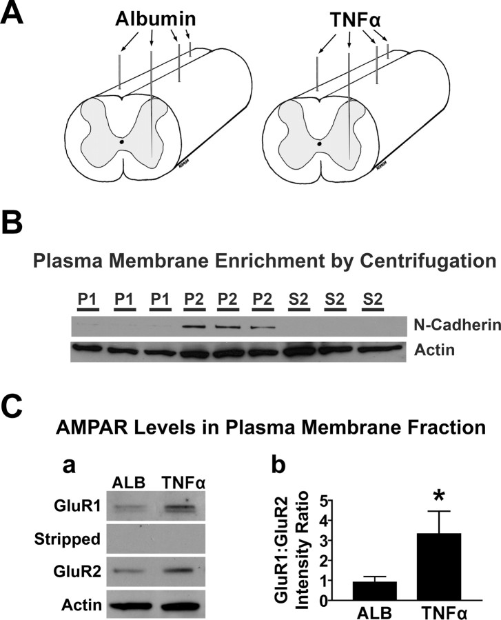
Biochemical evaluation of AMPAR trafficking to the plasma membrane after TNFα. A, Procedure used to inject the spinal gray matter with nanoliter quantities of TNFα or vehicle containing albumin. B, Subcellular fractionation by low speed centrifugation yields a modest enrichment in the plasma membrane protein N-cadherin in the P2 fraction (supplemental Fig. 1, available at www.jneurosci.org as supplemental material); however, actin is observed in all fractions. C, Densitometric analysis of Western blots from the P2 fraction reveals an increase of the GluR1:GluR2 ratio within the same blots after stripping and reprobing, suggesting a greater proportion of AMPARs that lack the GluR2 subunit in the plasma membrane after TNFα (*p < 0.05, Mann–Whitney U; n = 5 subjects per group); however, actin levels did not change (p > 0.05). Representative examples (C) reflect tissue from an albumin subject and a TNFα subject that were run on the same gel. Bars indicate group means of densitometry averaged across subjects. Error bars indicate SEM.

