Figure 6.
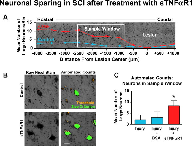
Sequestration of TNFα with sTNFαR1 saves motoneurons in the injured spinal cord. A, Representative tissue montage illustrating the sampling method and results of automated counts from treated (red) and pooled control (blue) conditions as a function of distance from lesion center. The sample window represents a fixed distance from the lesion center that was selected a priori to test for sparing in the lesion penumbra. B, Representative high power images depicting the number of Nissl-positive large neurons within the sample window after control or sTNFαR1 treatment. Cells included by the counting algorithm are highlighted in green. Scale bar, 40 μm. C, Group means for the automated cell counts within the sample window. (*p < 0.05 from control conditions, n = 4 subjects for sTNFαR1 and injury groups, and n = 3 subjects for BSA). Error bars indicate SEM.

