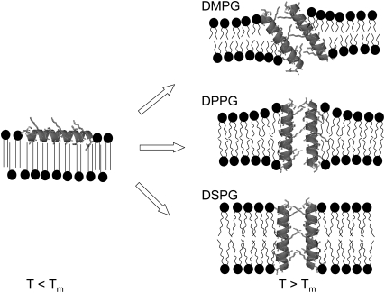
FIGURE 8.
Schematic representation of PGLa-associated structural changes in PG bilayers. Below the main phase transition (T < Tm), all three studied lipids exhibit a quasi-interdigitated phase in the presence of PGLa. The peptide aligns parallel to the membrane surface and its hydrophobic moieties are shielded by the hydrocarbon chains of the opposing lipid monolayer. In the fluid phase (T > Tm), above a critical threshold concentration, PGLa inserts into the PG membranes and most likely forms toroidal pores. In DMPG membranes, PGLa inserts at an angle and leads to a small increase in membrane thickness. The membrane thickening effect is most pronounced for DPPG membranes, where PGLa inserts vertically into the bilayer, and least pronounced for DSPG bilayers, in which no membrane deformation is necessary due to a match of the hydrophobic lipid core with the hydrophobic peptide length.

