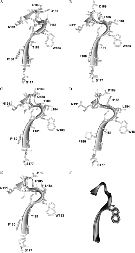
FIGURE 4.
Homology models showing the amino acid backbone of the loop B region of the 5-HT3 receptor. (A) Unbound structure (modeled from AChBP with no ligand, PDB ID 2byn). (B) Agonist bound, (modeled from AChBP with carbomylcholine, PDB ID 1uv6). (C) Agonist bound, (modeled from AChBP with epibatidine, PDB ID 2byq). (D) Large antagonist bound (modeled from AChBP with α-cobratoxin, PDB ID 1yi5). (E) Small antagonist bound, (modeled from AChBP with 2-methyllcaconitine, PDB ID 2byr) (F) Overlays of the modeled loop B backbones from A–E compiled with Swiss-PdbViewer “magic fit”, using residues Ser-177 to Asn-191 as a reference point.

