Figure 6.
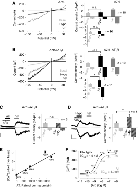
Expression of AT1R leads to mechanosensitivity of aortic SMCs. Whole-cell recordings from non-transfected (A) and A7r5 cells over-expressing AT1R (B–D). (A, B) IV relationships before ‘Basal', during hypotonic stimulation, ‘Hypo', and receptor stimulation with 1 μM vasopressin ‘VP' (left) and current density analyses at ±60 mV (right) are displayed. (C, D) Current time courses at ±60 mV with zero current levels (stippled lines); time scale bars 60 s and 100 pA (left) and current densities in the presence of neutralizing antibody, ‘nAb' (right) are displayed. (E) [Ca2+]i increases by hypotonic stimulation in HEK293 cells transfected with different amounts of AT1R-Venus cDNAs. Venus fluorescence was correlated with receptor densities. Numbers indicate the number of summarized cells and of independent transfections. (F) Concentration–response curves to AII of fura-2-loaded HEK293 cells stably expressing AT1R-Venus in isotonic and in hypotonic (273 mOsm kg−1) solutions.

