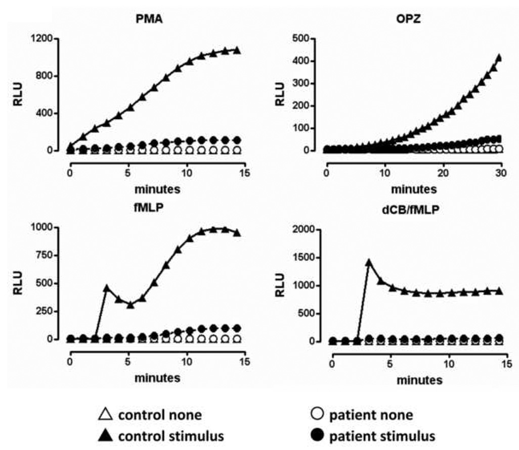
Figure 1. Evidence for neutrophil dysfunction.
Isolated neutrophils in HBSS/0.1% gelatin from the patient (circles) and a control donor (triangles) were incubated in the presence of 20µM luminol. The indicated stimulus (solid symbols) or buffer (None, open symbols) was added either directly (100nM PMA or 0.05mg/ml OPZ) or after established baseline readings (1µM fMLP or 10µM dCB + 1µM fMLP). Relative light units (RLU) were recorded continuously for 15 or 60 min depending on the stimulus. All stimuli were tested in duplicate wells and the patient’s cells were examined on two different occasions. Data shown are from one experiment.

