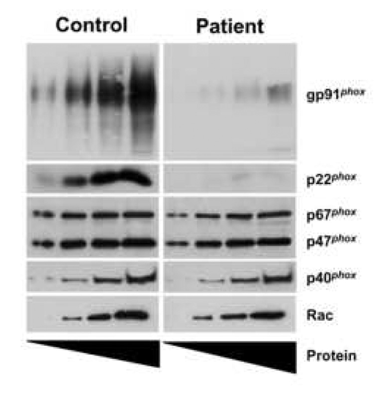
Figure 3. The patient expresses low levels of gp91phox and p22phox.
Increasing amounts of isolated neutrophil cytosolic and membrane fractions (1, 2.5, 5, 10µg for gp91phox; 5, 10, 20, 40µg for all other components) from both a control subject (left) and the patient (right) were prepared in Laemmli sample buffer, subjected to SDS-PAGE and transferred to nitrocellulose. Western blot analysis was performed on the membrane fractions for gp91phox and p22phox. Cytosolic fractions were probed for p67phox, p47phox, p40phox and Rac. The blots are representative of two experiments.

