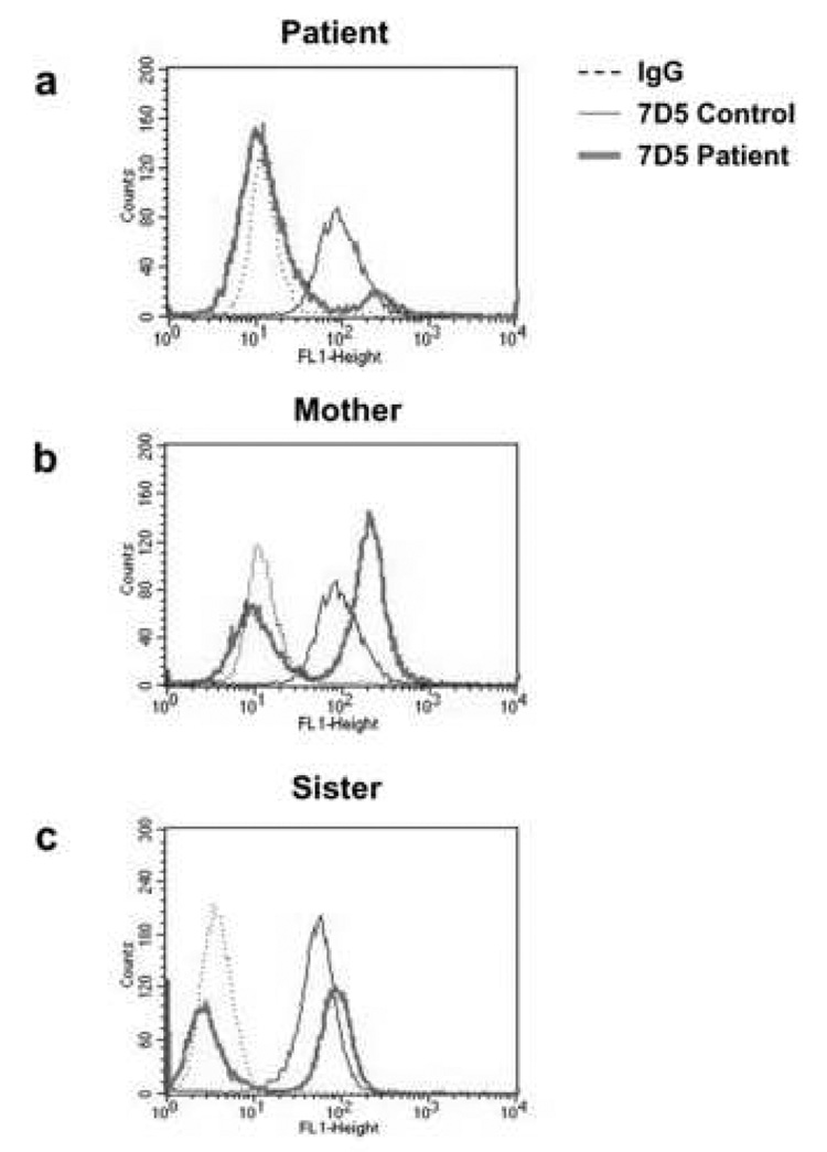
Figure 4. The patient, her mother, and sister are X-CGD carriers.
Isolated neutrophils (5×10⁶ cells/mL) from a control donor (thin solid line) or the indicated subject (thick solid line, 4a, Patient; 4b, Mother; 4c, Sister) were suspended in cold PBS/0.1% gelatin and stained with either irrelevant IgG (dashed lines) or mAb 7D5 (solid lines). Cells were then washed and incubated with FITC-goat anti-mouse secondary Ab and analyzed in a flow cytometer. Data are expressed as number of cells versus fluorescent intensity and are representative of at least three experiments.

