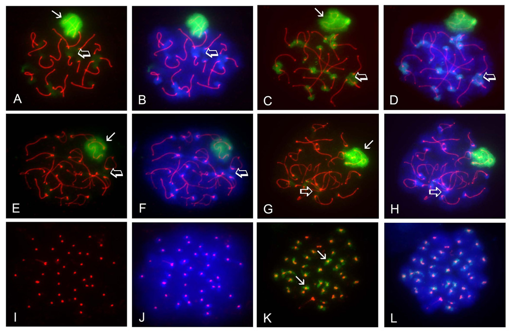
FIGURE 1. Both SUMO1 and SUMO2/3 accumulate in the XY body and heterochromatic chromocenters of pachytene and diplotene spermatocytes, but only SUMO2/3 localize to pericentromeric chromatin in metaphase I spermatocytes.
Surface-spread meiotic chromatin was immunolabeled with a rat polyclonal antibody specific for the synaptonemal complex marker SYCP3 (red), a mouse monoclonal antibody specific for SUMO1 (A, B, E, F, I, J) or a mouse monoclonal antibody specific to SUMO2/3 (C, D, G, H, K, L). Cells were counterstained with the fluorescent dye 4’, 6-diamidino-2-phenylindole (DAPI; blue) to visualize chromatin (B, D, F, H, L). A–B) Pachytene spermatocyte immunolabeled with the anti-SUMO1 antibody (green) showed localization of SUMO1 to the XY body (arrow) and chromocenters (open arrows). C–D) SUMO2/3 (green) also localize to the XY body (arrow) and chromocenters (open arrows) in pachytene spermatocytes. E–F) The SUMO1 signal (green) persists over the XY body (arrow) in diplotene spermatocytes, but diminishes at the chromocenters (open arrows). G–H) The SUMO2/3 signal (green) also persists in the XY body (arrow) in diplotene spermatocytes, but also diminishes at the chromocenters (open arrows). I–J) SUMO1 is not detected in meiosis I spermatocytes, as seen by the absence of green signal in these cells. Centromeres are identified by anti-SYCP3 labeling (red). K–L) SUMO2/3 (green) accumulate in the vicinity of sister centromeres (arrows) in MI spermatocytes. Magnification = 1000X.

