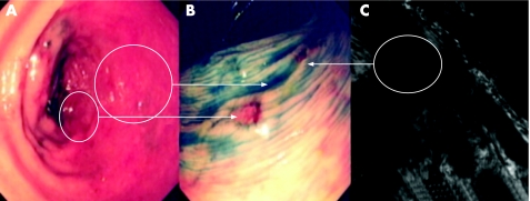
Figure 10 (A) Conventional “white light” views of the distal descending colon in a patient with longstanding pan‐colitis. There is focal erythema suggestive of a potential lesion. (B) Indigo carmine 0.5% chromoscopy reveals a well circumscribed flat elevated lesion with central depression (Paris class 0‐IIa+IIc). Detailed morphological assessment of this lesion indicates a high probability of submucosally invasive carcinoma. (C) A repeat colonoscopy using the confocal laser scanning endomicroscope shows a dilated and tortuous vascular net pattern at 60–80 μm in the z‐axis with complete destruction of the normal cryptic architecture. The lesion fulfils neoplastic criteria using Mainz the confocal imaging classification. Subsequent proctocolectomy was performed which revealed a stage T2/N1 well differentiated adenocarcinoma with associated lympho‐venous infiltration.

An official website of the United States government
Here's how you know
Official websites use .gov
A
.gov website belongs to an official
government organization in the United States.
Secure .gov websites use HTTPS
A lock (
) or https:// means you've safely
connected to the .gov website. Share sensitive
information only on official, secure websites.
