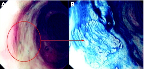
Figure 2 (A) Conventional “white light” endoscopic view of the distal sigmoid colon in a patient with longstanding pan‐colitis. There is focal mucosal pallor and nodularity. (B) Indigo carmine 0.5% chromoscopy shows a flat (Paris type 0‐IIb) lesion. The crypt pattern is a type I. On table diagnosis would favour a benign hyperplastic/metaplastic lesion. Endoscopic resection is therefore not indicated for this lesion.

An official website of the United States government
Here's how you know
Official websites use .gov
A
.gov website belongs to an official
government organization in the United States.
Secure .gov websites use HTTPS
A lock (
) or https:// means you've safely
connected to the .gov website. Share sensitive
information only on official, secure websites.
