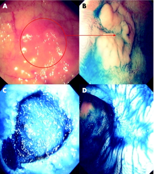
Figure 4 (A) Conventional “white light” view of the proximal sigmoid colon in a patient with distal colitis of >25 years duration. Note the focal mucosal erythema and loss of vascular net pattern. (B) Indigo carmine 0.5% chromoscopy clearly delineates a flat elevated circumscribed lesion with an area of central depression (Paris classification 0‐IIa+IIc). The adjacent mucosal architecture is within normal limits. The lesion endoscopically is an adenoma‐like mass (ALM). Endoluminal resection is indicated. (C) Endoscopic submucosal dissection using cap assistance has been performed. The lesion has been resected en bloc. Note the exposed underlying muscularis propria. (D) Post‐resection chromoscopic views of the lesion at 1 month. Note the depressed resection crater but complete re‐epitheliasation. There is no evidence of neoplastic crypt architecture indicating curative resection.

An official website of the United States government
Here's how you know
Official websites use .gov
A
.gov website belongs to an official
government organization in the United States.
Secure .gov websites use HTTPS
A lock (
) or https:// means you've safely
connected to the .gov website. Share sensitive
information only on official, secure websites.
