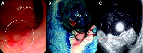
Figure 5 (A) Conventional “white light” view of the posterior‐lateral rectum in a patient with longstanding pan‐colitis. Note the focal nodularity and erythema. (B) Indigo carmine 0.5% chromoscopy delineated a flat elevated lesion with deep central depression (Paris class 0‐IIa + IIc). The lesion is highly suggestive of a submucosally invasive carcinoma. Endoscopic “through the scope” mini probe ultrasound is therefore used to locally stage the lesion. (C) 12.5 MHz mini probe “through the scope” ultrasound shows complete disruption of the third and fourth hypoechoic layers with infiltration of the muscularis. The lesion is a stage T2 carcinoma. Pan‐proctocolectomy is indicated.

An official website of the United States government
Here's how you know
Official websites use .gov
A
.gov website belongs to an official
government organization in the United States.
Secure .gov websites use HTTPS
A lock (
) or https:// means you've safely
connected to the .gov website. Share sensitive
information only on official, secure websites.
