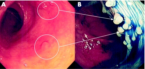
Figure 8 (A) Conventional “white light” views of the distal sigmoid colon in a patient with longstanding pan‐colitis. Note the focal mucosal pallor and nodularity of the mucosa. (B) Chromoscopy using 0.5% indigo carmine shows multiple sessile (Paris 0‐Is) lesions. The endoscopic diagnosis is most likely to be inflammatory polyps. Endoluminal resection is therefore not indicated. Further characterisation using high magnification imaging or confocal endomicroscopy would be beneficial.

An official website of the United States government
Here's how you know
Official websites use .gov
A
.gov website belongs to an official
government organization in the United States.
Secure .gov websites use HTTPS
A lock (
) or https:// means you've safely
connected to the .gov website. Share sensitive
information only on official, secure websites.
