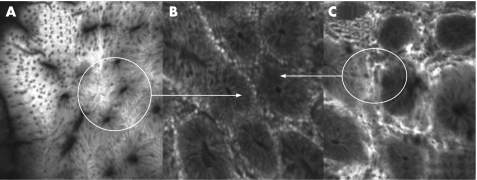
Figure 9 (A) Superficial crypt architecture of the suspected inflammatory lesion shown in fig 8 using the Pentax confocal endomicroscope and intravenous fluoroscene. The crypt architecture at 10 μm z‐axis penetration is organised but with crypt elongation. There is no goblet cell depletion indicating an inflammatory aetiology. (B) Confocal laser scanning imaging at 100 μm in the z‐axis. The crypts are well aligned in a concentric architecture again indicating a non‐neoplastic nature. (C) Deep 250 μm laser scanning images shows a hexagonal per‐cryptic vascular net pattern with no extravasation of fluoroscene. The “black dots” represent individual red cells within the per‐cryptic capillary. This vascular pattern according to Mainz criteria is suggestive of hyperplasia or normal colonic mucosa.

An official website of the United States government
Here's how you know
Official websites use .gov
A
.gov website belongs to an official
government organization in the United States.
Secure .gov websites use HTTPS
A lock (
) or https:// means you've safely
connected to the .gov website. Share sensitive
information only on official, secure websites.
