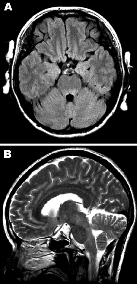
Figure 1.
Magnetic resonance images of the brain. A) Hyperintense lesions in the tegmentum of the pons in the axial section of the fluid-attenuated inversion recovery image. B) In the sagittal section of the T2-weighted image, hyperintense lesions are present in the tegmentum of the midbrain, pons, and medulla oblongata.

