Figure.
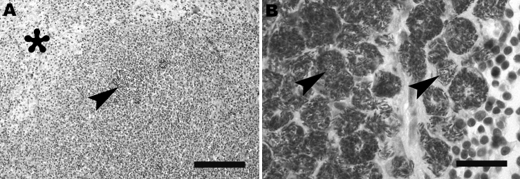
A) Mesenteric lymph node of Yorkshire Terrier shows diffuse granulomatous lymphadenitis with extensive infiltration of macrophages, foci of pyogranulomatous inflammation (arrowhead), and focal necrosis (asterisk). Hematoxylin and eosin stain; scale bar represents 100 μm. B) Retropharyngeal lymph node of schnauzer shows innumerable acid-fast bacilli (arrows) within the cytoplasm of macrophages. Ziehl-Neelsen stain; scale bar represents 25 μm.

