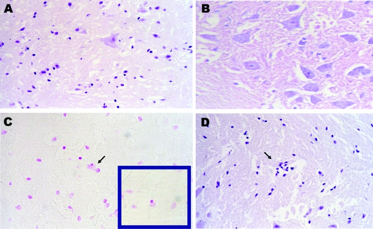
Figure 2.
Pathologic images obtained from a carcass of Eptesicus isabellinus. The bat was captured while flying but died during handling. Brain specimen was positive for lyssavirus antigens by immunofluorescence and for European bat lyssavirus 1 RNA by reverse transcription–PCR. A) Neural degeneration in brain by hematoxylin and eosin stain (H&E); magnification ×400. B) Negative Seller stain in spinal cord indicating the absence of Negri bodies; magnification ×400. C) Positive Feulgen reaction in brain, glial cell neurophagia; magnification ×200. D) Focal proliferation of glial cells by H&E; magnification ×100.

