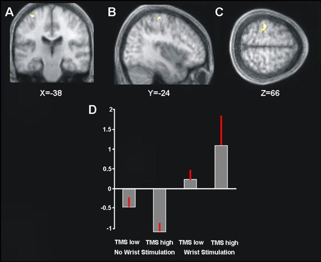
Figure 2.
A–C, Areas showing an interaction between high- minus low-intensity right parietal TMS, and the presence minus absence of right median nerve sensory stimulation, within regions responding to right median nerve stimulation overall (compare Fig. 1). Significant (p < 0.05, FWE corrected) interaction effects are shown on coronal (A), sagittal (B), and transverse (C) slices of the averaged structural scans. The left SI is clearly implicated. D, Plot of the mean percentage of BOLD signal change extracted from the left SI cluster of the interaction contrast shown in A–C. High- versus low-intensity TMS over right parietal cortex increased the BOLD signal for left SI during right median nerve stimulation (compare third and fourth bars), but the same high TMS decreased the BOLD signal for left SI in the absence of somatosensory input (compare first and second bars). Restated, high-intensity right parietal TMS increased the differential response of left SI to the presence versus absence of right-hand stimulation (compare fourth and second bars in the histogram), compared with low-intensity TMS (compare third and first bars).

