Figure 3.
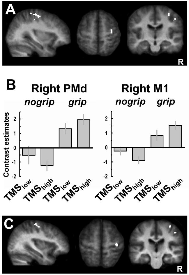
Interaction between TMS intensity and current motor state for BOLD signals. A. SPMs showing a significant interaction [(TMShighnogrip - TMSlownogrip) - (TMShigh-grip - TMSlow-grip)] in right PMd contralateral to TMS stimulation (x,y,z: 34, -16, 58; assigned to Area 6 with a 60% probability, [Eickhoff and others 2006]) and right M1 (36, -26, 62; assigned to Area 4a with a 60% probability). These same regions were also modulated by the grip task (i.e. were active during left-hand grip), see Table 1, Figure 1C.
B. SPM parameter estimates showing that TMShigh to left PMd at rest led to a relative activity decrease in contralateral PMd and M1 (BA4a), as compared to TMSlow. By contrast, when applied during active left-hand grip, TMShigh to left PMd now led instead to a relative increase in activity in these regions. This illustrates the state-dependent influence of left PMD TMS on contralateral motor areas which are implicated in hand movement control.
C. Regions implicated in the grip task that also show changes in functional coupling (as revealed by an independent psychophysiological interaction analysis) with the site of TMS stimulation (left PMd), as a function of event-related condition. A significant context-dependent covariation with left (stimulated) PMd was revealed in two similar clusters as for the SPM interaction analysis, located in contralateral right PMd (x, y, z = 36, -14, 56) and right M1 (x, y, z = 36, -24, 64). Thus, the functional coupling between left PMd and right PMd and right M1 was stronger for TMShigh versus TMSlow during grip than during no-grip. Results are projected onto the average normalized structural scans of all participants (random effects analysis, corrected for multiple comparisons across the whole brain at T>4 and extent (cluster) threshold of P < 0.05). R = right.

