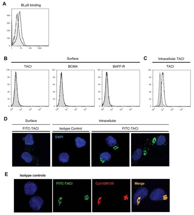
FIGURE 10. BLyS receptor expression.

A. Soluble BLyS binding was determined by flow cytometry. DCs were incubated with (open histogram) or without (filled histogram) 2 μg/ml soluble BLyS for 2 h at 37°C, washed, incubated with biotinylated anti-BLyS antibody, and stained with streptavidin-conjugated PE. Isotype control was indicated by the dotted histogram. B. Surface staining of TACI, BCMA, and BAFF-R. C. Intracellular TACI staining. Specific receptor expression was indicated by the open histograms and isotype and fluorescence controls were indicated by the filled histograms. D. Subcellular localization of TACI in DCs by confocal immunofluorescence microscopy. Surface and intracellular TACI expression was assessed using a TACI specific goat antibody. E. Cells were costained with anti-TACI and anti-GM130 antibodies. Colocalization was confirmed when the images were merged (right panel). Isotype matched control primary antibody staining is also shown in D and E.
