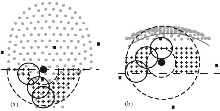
Figure 1.
Example of model origin search for vertex orientation; grid slices through the fMCG origin (‘+’, 2 cm spacing) superimposed onto sensor array projection (gray dots) with estimated fMCG source location (large filled circle, r = 1.5 cm), 3 candidate model spheres (large open circles, r = 4.5 cm), and abdominal localization coils (filled squares). The dashed concentric circles (r = 6 and 15 cm) define the annular shell about the heart center for grid constraint. (a) projection onto x-y plane; the horizontal dashed line limits the grid to head origins below the heart in this example, (b) projection onto x-z plane; the horizontal dashed line limits the grid depth (4.5 cm deeper than MCG center). The gray arc estimates the inner maternal abdominal surface (3 cm from the sensor coils).

