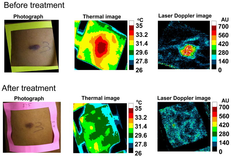
Figure 1.
Typical example of light photography, thermal imaging, and laser Doppler imaging of a KS lesion prior to therapy and after therapy. As can be seen, the thermal and laser Doppler images normalized after treatment, while the lesion was still evident by light photography. Reproduced with permission from Hassan et al., Technology in Cancer Research and Treatment, 3 (5), 451–7, 2004.

