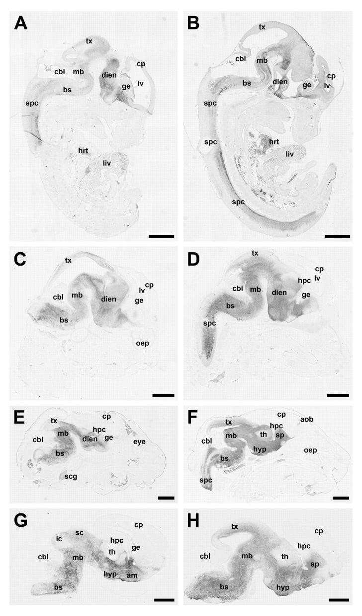
Figure 3.
Representative photomicrographs demonstrate the expression pattern of Ahi1 in the developing mouse at embryonic day (E) 12.5 (A,B), E14.5 (C,D), E16.5 (E,F) and E18.5 (G,H) in lateral sections through the brain (A,C,E,G) and in midsagittal sections through the brain (B,D,F,H). Ahi1 expression is most pronounced in the ventral aspects of the developing brain (developing amygdala, hypothalamus, midbrain, brainstem, and spinal cord) with no detectable immunoreactivity in the developing cortical plate or cerebellum. Bar indicates 1 mm. am: amygdala; aob: accessory olfactory bulb; bs: brainstem; cbl: cerebellum; cp: cortical plate; dien: diencephalon; ge: ganglionic eminences; hpc: hippocampus; hyp: hypothalamus; ic: inferior colliculus; lv: lateral ventricle; mb: midbrain; oep: olfactory epithelium; sc: superior colliculus; spc: spinal cord; scg: superior cervical ganglion; sp: septal nucleus; th: thalamus; tx: tectum

