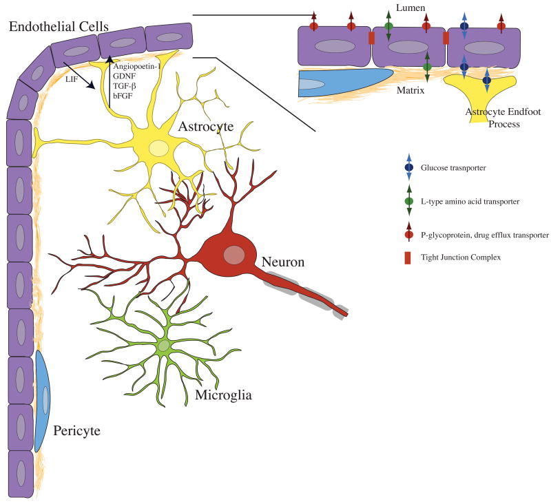
Figure 1. Endothelial cells interact with a variety of brain cell types to produce specialized blood-brain barrier tissue.
Astrocytes produce several factors that have been shown to induce formation of tight junctions, and expression of blood-brain barrier specific protein markers. Factors including angiopoetin-1, GDNF, TGFβ and bFGF can activate endothelial cells by modulation of intracellular cAMP levels. Endothelial cells signal astrocytes via leukemia inhibitory factor (LIF). The chemical signaling among neurons, microglia, and pericytes is not as well understood. However, neurons are metabolically coupled to both astrocytes and vascular endothelial cells, and are rarely found more than 20–30 micrometers from capillary microvessels. Microglia are often associated with vascular elements in tissue slices, and pericytes occupy space within the basal lamina, directly associating with the abluminal surface of endothelial cells. Inset. Astrocytes closely associate with endothelial cells by contacting the basal lamina with specialized endfoot processes. Barrier function is determined by several factors, including tight junctions between endothelial cells, as well as cellular metabolism and transporter expression. Endogenous transporters of glucose and large neutral amino acids are present on the luminal and abluminal surfaces of endothelial cells. Xenobiotic efflux transporters such as p-glycoprotein (Pgp) are often located exclusively on the lumen-facing membrane of endothelial cells.

