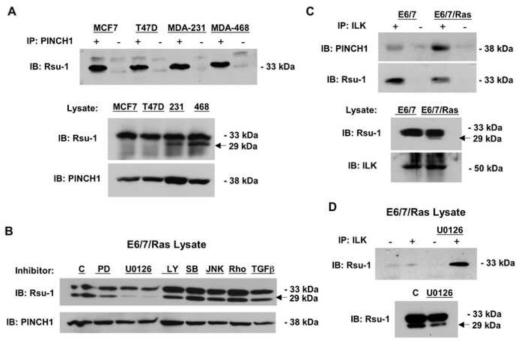
Fig. 2. The association of Rsu-1 with the PINCH1-ILK complex is altered in tumor cell lines.
(A) Lysates of human breast cancer cell lines were immunoprecipitated with anti-PINCH1. The immunoprecipitates were analyzed by Western blotting for co-immunoprecipitation of endogenous Rsu-1. Only p33 Rsu-1 is detected in PINCH1 immunoprecipitates. (B) The Ras-transformed immortalized human astrocytes were treated with inhibitors at the indicated concentrations for 36 h and the effect on p33 and p29 Rsu-1 protein expression was determined by Western blotting. Inhibitors: 10 µM PD98059, 10 and 20 µM U0126 (left and right lanes, respectively), 10 µM LY29402, 500 nM SB20350, 100 nM JNKII (SP600125), 100 nM RhoK inhibitor (Y27632), 5 ng/ml TGFβ. (C) Lysates of immortalized human astrocytes and the Ras-transformed astrocyte cell line were immunoprecipitated with anti-ILK. The immunoprecipitates were analyzed by Western blotting for co-immunoprecipitation of endogenous Rsu-1 and PINCH1. (D) The ILK-immunoprecipitates of Ras-transformed astrocytes were analyzed by Western blotting for co-immunoprecipitation of endogenous Rsu-1 with and without pre-treatment of the cells with U0126.

