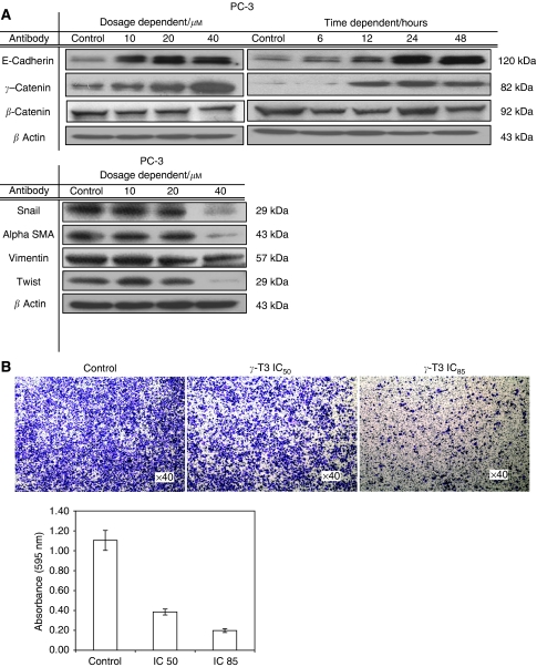
Figure 4.
Inhibition of cell invasion by γ-T3 treatment. (A) 24-h dose-dependent and IC50 time-dependent γ-T3 treatment induces the expression of epithelial markers (E-cadherin, γ-catenin), but suppresses the expression of mesenchymal markers (vimentin, twist and α-SMA) and E-cadherin's repressor (snail). (B) The invasive androgen-independent PCa cells (PC-3) treated with the indicated dosage of γ-T3 was harvested and then plated into the Matrigel-coated (0.5 mg ml−1) insert. Cells invaded through the membrane were stained with crystal violet and the images were photographed under a microscope. After being lysed with extraction buffer, intensity at 595 nm was measured.

