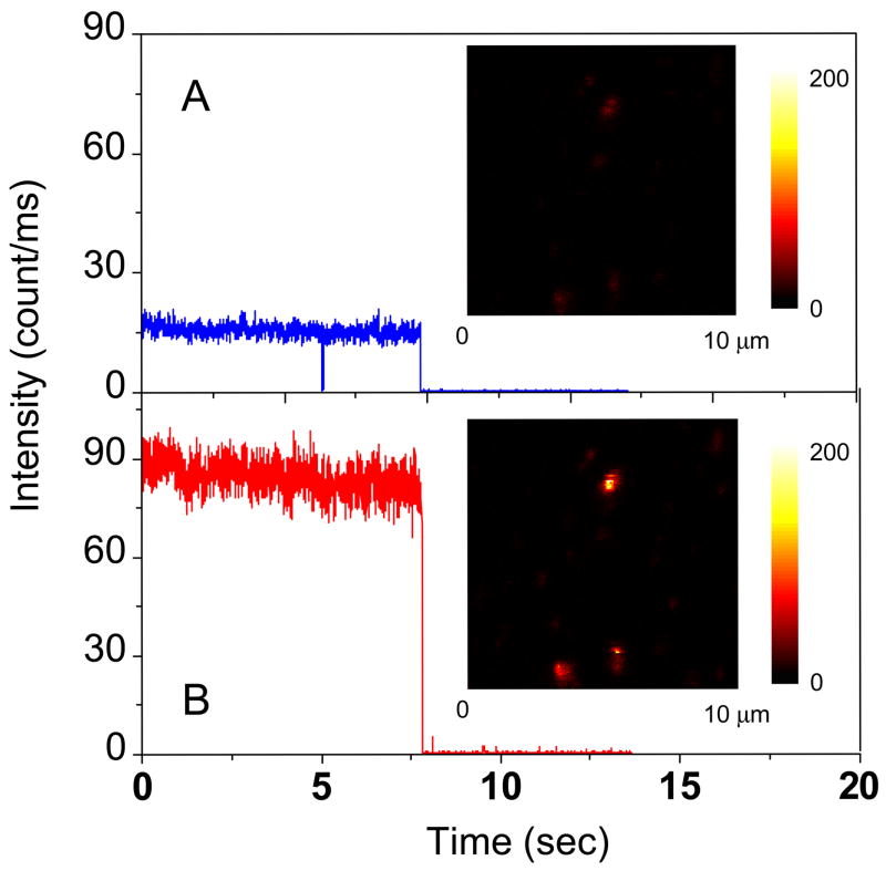
Figure 6.
Respective time-trace intensities of a donor-acceptor pair bound on the silver particle as seen in (a) the donor channel or (b) the acceptor channel. The insets represent the respective fluorescence images. The images are 150 × 150 pixels, with in integration time of 0.6 ms per pixel. Both images were recorded simultaneously using two SPAD detectors.

