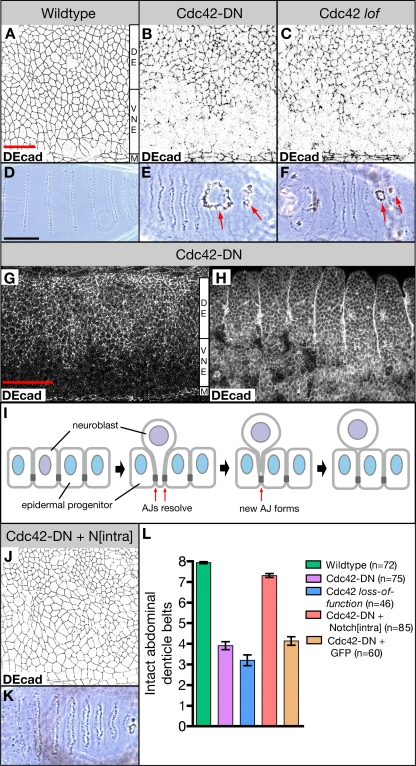
Figure 1.
Ventral ectodermal defects in embryos overexpressing Cdc42-DN and Cdc42 loss-of-function embryos. (A–C) Wild-type embryo (A), embryo overexpressing Cdc42-DN under the control of da-Gal4 (Cdc42-DN; B), and embryo produced by a Cdc423/Cdc426 female (Cdc42 lof; C) labeled for the AJ marker DEcad. (D–F) Ventral cuticle of wild-type embryo (D), Cdc42-DN embryo (E), and Cdc42 loss-of-function embryo (F). Arrows indicate holes on the ventral surface. (G and H) Cdc42-DN embryos labeled for DEcad at stages 11 (G) and 14 (H). (I) Illustration of neuroblast ingression. As a neuroblast (purple) begins to ingress from the ventral neuroectoderm, existing AJs (dark gray) at the neuroblast/epidermal progenitor (blue) boundaries resolve. New AJs form between neighboring epidermal cells. (J and K) DEcad stain (J) and ventral cuticle (K) of embryos expressing both Cdc42-DN and Nintra under the control of da-Gal4. (L) The extent of ventral cuticle defects was quantified by counting the number of intact abdominal denticle belts (mean ± SEM [error bars]). The difference in the number of intact belts is highly significant (P < 0.001) for Cdc42-DN embryos versus wild type, Cdc42 loss-of-function embryos versus wild type, and Cdc42-DN Nintra embryos versus Cdc42-DN embryos. Coexpression of GFP with Cdc42-DN under the control of da-Gal4 did not affect the severity of ventral cuticle defects caused by Cdc42-DN. M, ventral midline; VNE, ventral neuroectoderm; DE, dorsal ectoderm. Bars: (A–C and J) 20 μm; (D–F and K) 100 μm; (G and H) 50 μm.

