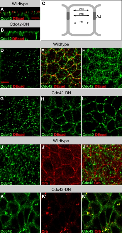
Figure 3.
Localization of Cdc42 in epithelia of wild-type embryos and embryos expressing Cdc42-DN. (A and B) XZ reconstruction of image stacks of neuroectodermal cells of wild-type (A) and Cdc42-DN (B) embryos labeled for DEcad and Cdc42. (C) Schematic of an epithelial cell indicating the focal planes shown in panels D–I. (D–I) Apical membrane (D and G), AJ (E and H), and basolateral membrane (F and I) single-plane views of wild-type (D–F) and Cdc42-DN (G–I) embryos labeled for DEcad and Cdc42. (J and K) Wild-type (J) and Cdc42-DN (K) embryos labeled for Crb and Cdc42. Bars, 5 μm.

