Figure 4.
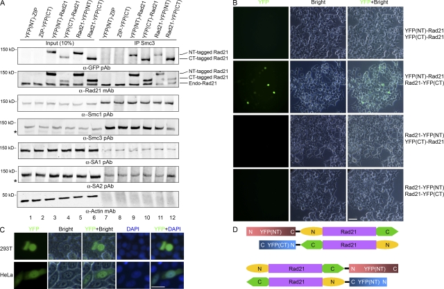
Fluorescent protein fragment complementation assay showing the Rad21–Rad21 interaction and the antiparallel orientation. (A) YFP(NT) or YFP(CT) were fused to either the NT or CT end of Rad21. YFP-fused Rad21 constructs were expressed in 293T cells (lanes 3–6), and their interaction with the cohesin complex was examined by IP of the endogenous Smc3 using rabbit anti-Smc3 antisera (lanes 9–12). *, nonspecific band. (B) 293T cells were cotransfected with YFP(NT)- and YFP(CT)-fused Rad21 plasmids (a total of four combinations). YFP fluorescence was examined under a fluorescent microscope 40 h after transfection. (C) YFP fluorescence–positive 293T and HeLa cells transfected with the combination of YFP(NT)-Rad21 and Rad21-YFP(CT) at 400× magnification. (D) Possible antiparallel orientation of Rad21–Rad21 interactions. Only the combination of plasmids in the top panel results in the fluorescence. Bars, 25 μm.

