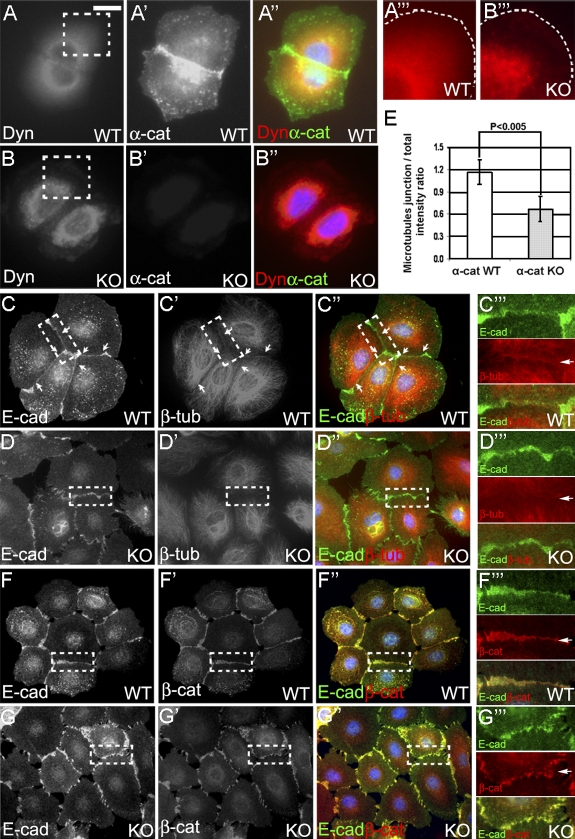
Figure 2.
α–E-catenin is necessary to extend dynactin and the microtubule cytoskeleton to the cell periphery and localize microtubules to the AJs. (A and B) Immunofluorescence staining of wild-type (WT) and α–E-catenin−/− (KO) cells with antidynamitin (Dyn) and anti–α-catenin (α-cat) antibodies. Regions in dashed boxes are shown at higher magnifications in A‴ and B‴. The cell edges are outlined with white dashed lines. (C and D) Immunofluorescence staining of wild-type and α–E-catenin−/− cells with anti–E-cadherin (E-cad) and anti–β-tubulin (β-tub) antibodies. Regions containing cell–cell junctions (dashed boxes) are shown at higher magnifications in C‴ and D‴. (E) Quantitation of microtubule accumulation at cell–cell junctions. Pairs of contacting cells displaying accumulation of E-cadherin at cell–cell borders were randomly selected. The levels of cell border accumulation of microtubules are expressed as ratios of the mean β-tubulin staining intensity at cell–cell junctions over the mean total β-tubulin intensity within two contacting cells. Each bar represents the mean value; n = 50. The p-value was determined by a t test. The error bars represent standard deviation. (F and G) β-Catenin localizes to AJs in α–E-catenin−/− cells. Immunofluorescence staining of wild-type and α–E-catenin−/− cells with anti–E-cadherin and anti–β-catenin (β-cat) antibodies. Regions containing cell–cell junctions (dashed boxes) are shown at higher magnifications in F‴ and G‴. Arrows denote the positions of the AJs. Bars: (A–A″ and B–B″) 16 μm; (A‴ and B‴) 8 μm; (C–C″, D–D″, F–F″, and G–G″) 30 μm; (C‴, D‴, F‴, and G‴) 13 μm.

