Figure 5.
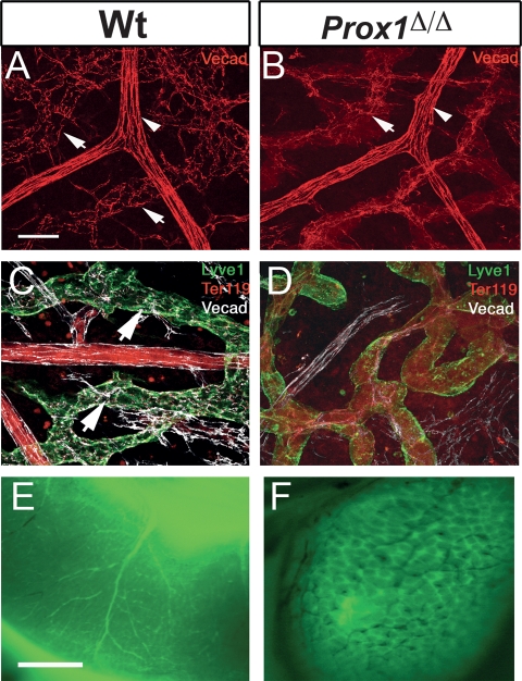
Prox1 mutant vessels exhibit disrupted cell adhesion properties and aberrant connections between the lymphatic vasculature and blood vasculature. (A) VE-cadherin expression was found in button-like junctions between LECs of submucosal lymphatics of P5 wild-type intestines (arrows). Strong uniform expression was seen in blood vessels (arrowhead). (B) In contrast, VE-cadherin staining was weak or absent in the lymphatics of conditional mutant littermates (arrow). (C) VE-cadherin (white) expression was found in normal button-like junctions between the LECs of the Lyve1+ (green) submucosal lymphatics of P5 wild-type intestines (arrows). (D) In contrast, VE-cadherin staining was weak or absent in the lymphatics of conditional mutant littermates, where Lyve1 expression level was down-regulated. Ter119 (red) staining within the conditional mutant lymphatic also indicated the presence of red blood cells. (E) Cardiac perfusion of FITC-dextran labeled the blood vasculature of the P5 wild-type intestine. (F) A similar assay performed on P5 Prox1Δ/Δ littermates labeled not only the blood vasculature but also all of the submucosal lymphatics. The black small vessels seen in this panel correspond to blood-filled small capillaries that were not perfused with the PFA/FITC-dextran solution. Bars: A,B, 50 μm; E,F, 1 mm.

