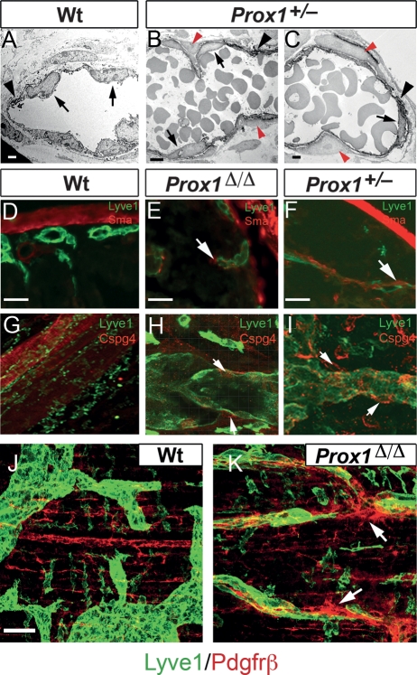
Figure 6.
Prox1 mutant pups display ectopic pericyte investment. (A) Electron microscopic imaging of DAB Lyve1-stained (arrowhead) wild-type submucosal lymphatics exhibiting their typical morphology, ECs are indicated by the arrows. Images from Prox1+/− (B,C) pups revealed the abnormal presence of Lyve1 pericytes (red arrowheads) around some mutant lymphatic vessels. Wild-type lymphatics were devoid of Sma+ (D), Cspg4+ (G), and Pdgfrβ+ (J) pericytes. (E,F,H,I,K) Abnormal ectopic expression of Sma, Cspg4, and PDGFRβ (arrows) was detected in some of the lymphatics of the conditional mutant and Prox1+/− pups. Bars: A, 4 μm; B, 8 μm; C, 3 μm; D–I, 50 μm; J,K, 50 μm.

