Figure 4.
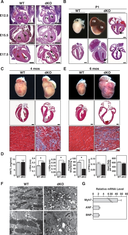
Abnormalities of embryonic and adult dKO mutant hearts. (A) Sections of wild-type and dKO hearts during embryogenesis. At E12.5, E15.5, and E17.5, dKO hearts were normal, except for dilatation of the RV and thinning of the RV-free wall. Arrowhead indicates VSD near the atrioventricular valve. (rv) Right ventricle; (lv) left ventricle. Bar, 500 μm. (B) Whole hearts and longitudinal sections of wild-type and dKO hearts at P1. Arrows point to VSD at the apex of heart and arrowhead to VSD near atrioventricular valve. Sections of three different dKO hearts are shown. (ra) Right atrium; (la) left atrium. Bar, 500 μm. (C) Hearts of wild-type and dKO mutant mice at 4 mo of age. Whole-mount pictures of the hearts are shown in the top panel. The middle panels show histological sections stained with Masson’s trichrome. The bottom panels show the interventricular septum at high magnification. Note extensive fibrosis of dKO heart, especially at the junction of the interventricular septum, where VSDs were frequently observed. (Middle panel) Bar, 1 mm. (Bottom panel) Bar, 20 μm. (D) Analyses of cardiac function by echocardiography. Four-month-old male miR-133a dKO mice and their control littermates (n = 11 for each group) were analyzed. (HW/TL) Heart weight-to-tibia length ratio; (LVIDd) left ventricular internal diameter at end-diastole; (LVIDs) left ventricular internal diameter at end-systole; (LVPWd) left ventricle posterior wall thickness at end-diastole. Asterisks indicate statistical significance. The P-values for the following measurements are HW/TL: P = 0.7684; LVIDd: P = 0.0065; LVIDs: P = 3.9e-007; fractional shortening: P = 2.0e-011; LVPWd: P = 0.5424; heart rate: P = 0.1102. (E) Hearts of wild-type and dKO mutant mice that died suddenly at 6 mo of age. Whole-mount pictures and Masson’s trichrome-stained sections of hearts of wild-type and dKO mice at the time of death are shown. The bottom panels show the interventricular septum at high magnification. Note severe ventricular dilatation and fibrosis of dKO hearts. (Middle panel) Bar, 1 mm. (Bottom panel) Bar, 20 μm. (F) Transmission electron micrographs of adult wild-type and dKO mutant mice at 4 mo of age show disorganized sarcomeres and mitochondrial abnormalities in the mutant. Arrowheads point to abnormal Z-lines and arrows point to mitochondria in dKO mutant heart. (Top panels) Bar, 2 μm. (Bottom panel) Bar, 1 μm. (G) Transcripts for the indicated markers of cardiac stress were measured by real-time PCR in RNA samples from wild-type and dKO mice at 4 mo of age. Expression levels in dKO mice are expressed relative to expression in wild-type mice (n = 3 for each genotype). Error bars indicate the SEM.

