Figure 5.
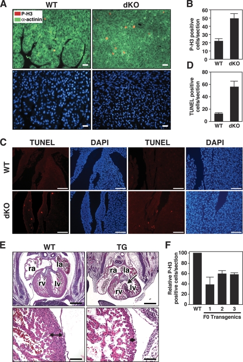
Abnormal cardiomyocyte proliferation and apoptosis in miR-133a dKO hearts. (A) Immunohistochemistry on heart sections of wild-type and dKO mutant mice at P1. Phospho-histone H3 (red), α-actinin (green), and Hoechst (blue) staining at 40× magnification show increased proliferation in the cardiomyocytes in dKO mutant mice. Bar, 20 μm. (B) Quantification of phospho-histone H3-positive cells was performed on three sections from each heart and averaged from six individual hearts. Error bars indicate the SEM. (C) TUNEL staining of wild-type and dKO hearts at P1 showed increased apoptosis in dKO mice. TUNEL-positive cells are located near the base (left panels) and apex (right panels) of the heart in dKO mice. DAPI staining indicates nuclei. Bar, 100 μm. (D) TUNEL-positive cells were quantified on multiple sections from each dKO heart. Error bars indicate the SEM. (E) Transgenic overexpression of miR-133a blocks proliferation of cardiomyocytes in vivo. Histological sections of wild-type and βMHC-miR-133a transgenic hearts at E13.5 are shown. The arrow in the top right panel points to a VSD. The bottom panels show higher magnifications of the left ventricular myocardium, which is about eight cells thick in wild type and only two cells thick in the transgenic. (Top panel) Bar, 500 μm. (Bottom panel) Bar, 100 μm. (F) Quantification of phospho-histone H3-positive cells in histological sections of hearts from wild-type and three independent F0 βMHC-miR-133a transgenic hearts at E13.5. Phospho-histone H3-positive cells were counted on three sections for each transgenic heart and normalized to the number of phospho-histone H3-positive cells of wild-type littermates. Error bars indicate the SEM.

