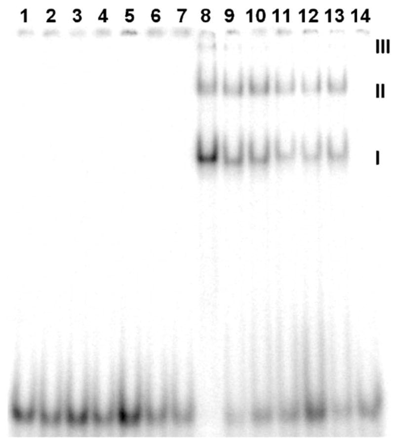
Fig. 4.

EMSA on wt bZIP bound to AP-1, C/EBP, XRE1, Partial site, E-box, and HRE. Each lane contains ~3000 cpm 32P-endlabeled 24-mer duplex. Lanes 1–7: free DNA. Lanes 8–14: DNA in the presence of 500 nM wt bZIP. Lanes 1 and 8, AP-1; lanes 2 and 9, C/EBP; lanes 3 and 10, XRE1; lanes 4 and 11, E-box; lanes 5 and 12, HRE; lanes 6 and 13, Partial site; lanes 7 and 14, nonspecific control. EMSA was run at 120 V for 4 h. I indicates bandshift from dimeric wt bZIP complexation. II and III indicate wt bZIP aggregates that bind to the duplexes and cause bandshifts (see text for discussion).
