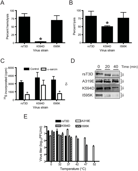
Figure 3. K594D exhibits altered capacity for membrane penetration.
(A) A 3% v/v solution of bovine erythrocytes was incubated with 5.4×10¹⁰ ISVPs of rsT3D or the indicated ϕ mutant at 37°C for 1 h. Hemolysis was quantified by determining absorbance of the supernatant at 415 nm. Hemolysis following treatment of an equal number of cells with virion-storage buffer or virion-storage buffer containing 1% TX-100 was considered to be 0 or 100%, respectively. Results are expressed as mean percent hemolysis for triplicate samples. Error bars indicate SD. *, P<0.05 as determined by Student's t-test in comparison to rsT3D. (B) L929 cells preincubated with 51Cr-labeled sodium chromate were adsorbed with 105 ISVPs/cell of rsT3D or the indicated ϕ mutant at 4°C for 1 h and incubated at 37°C for 4 h following addition of complete medium. The amount of 51Cr released into the medium was determined by liquid scintillation. 51Cr release following treatment of an equal number of cells with virion-storage buffer or by addition of 4% TX-100 to the medium was considered to be 0 or 100%, respectively. Results are expressed as mean percent lysis for triplicate samples. Error bars indicate SD. *, P<0.05 as determined by Student's t-test in comparison to rsT3D. (C) HeLa cells starved of cysteine and methionine were adsorbed with 106 ISVPs/cell of rsT3D or the indicated ϕ mutant at 4°C for 1 h. Infection was initiated in medium containing 35S-labeled cysteine and methionine in the presence or absence of α-sarcin. Cells were lysed following incubation at 37°C for 1 h. Proteins were precipitated with TCA, and acid-precipitable radioactivity was quantified by scintillation counting. Results are expressed as mean 35S incorporated for triplicate samples. Error bars indicate SD. *, P<0.05 as determined by Student's t-test in comparison to 35S incorporated in the absence of α-sarcin. (D) CsCl-treated ISVPs of rsT3D or the indicated μ1 mutant were incubated with trypsin at 4°C for the intervals shown. Samples were resolved by SDS-PAGE and immunoblotted using a MAb specific for μ1. The position of the δ band is shown. (E) ISVPs of rsT3D or the indicated μ1 mutant were incubated at the temperatures shown for 15 min. Residual infectivity was assessed by plaque assay. Results are expressed as mean residual titer for triplicate samples. Error bars indicate SD.

