
Fig. 2.
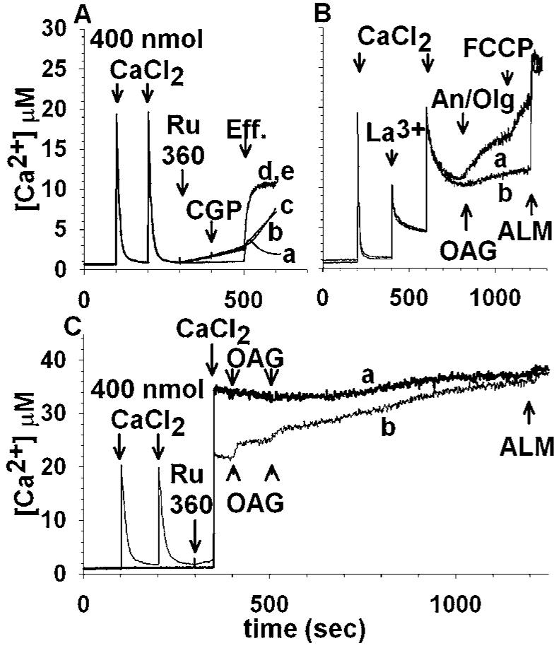
OAG-induced Ca2+ efflux from rat brain mitochondria is not mediated by known Ca2+ uptake and release pathways. (A): Mitochondria are loaded by exogenously added Ca2+ (each pulse is 400 nmol CaCl2) and the following compounds are administered: trace a, OAG (100 μM) at 500 s; trace b, 165 nM Ru360 at 300 s, 1 μM antimycin A3 plus 2 μM oligomycin at 500 s; trace c, 165 nM Ru360 at 300 s, 0.5 μM FCCP at 500 s; trace d, 165 nM Ru360 at 300 s, 100 μM OAG at 500 s; trace e, 165 nM Ru360 at 300 s, 20 μM CGP-37157 at 400 s, 100 μM OAG at 500 s; traces d and e are superimposed. “Eff.”: effectors. (B): 1 mM LaCl3 abolishes OAG-induced Ca2+ efflux, without affecting the reverse function of the uniporter: mitochondria are treated with 400 nmol CaCl2 at 350 and 600 s and 1 mM LaCl3 in between (500 s). Subsequently 1 μM antimycin A3 plus 2 μM oligomycin are administered at 800 s (trace a) or 100 μM OAG at 800 s (trace b). In trace a 0.5 μM FCCP was also added at 1100 s. In both traces, alamethicin (40 μg/ml) is added at 1200 s. (C): In trace a, rat brain mitochondria are not preloaded with Ca2+, and Ru360 (165 nM) is added at 300 s; subsequently, 800 nmol of CaCl2 is added at 350 s, followed by two pulses of OAG (100 μM each) at 400 and 500 s. In trace b, mitochondria are preloaded with two pulses of exogenously added CaCl2 (100 and 200 s, 400 nmol each); 165 nM Ru360 is added at 300 s, followed by an additional pulse of 400 nmol CaCl2 at 350 s. Subsequently OAG (two pulses of 100 μM each) are added at 400 and 500 s. In both traces, alamethicin (40 μg/mL) is added at 1200 s.