
FIG. 1.
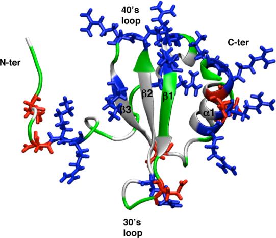
The structure and location of charged residues (shown in line form) in the canonical chemokine fold of hLtn (averaged NMR structure from PDB code 1J9O). The residues are colored based on the residue type: white for non-polar, green for polar, red for negatively charged and blue for positively charged residues.