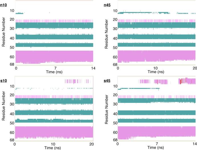
FIG. 3.
Secondary structures of hLtn during the four MD simulations under different conditions (see Table I for details). The color coding is: green for β strand, magenta for α helix, red for π-helix, white for turn and for coil. These plots are produced by VMD73 using the STRIDE algorithm for secondary structure determination72.

