
Figure 1.
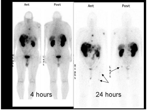
Planar 111In pentreotide single photon scintigraphy of a patient with metastatic carcinoid. Note the metastases in the right pelvis and femur on the 24-hour images that were not well visualized on the 4-hour image. The additional delay allows for improved blood pool clearance and tumor uptake.