
Figure 2.
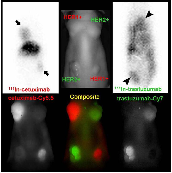
This image shows concordance between fluorescent optical and 111In gamma scintigraphy of HER2 and HER1 labeled antibodies. Optical imaging is a powerful pre-clinical tool; however it is difficult to image deep tissues. Gamma scintigraphy will be more suitable for clinical translation. Top row: (left and right) 50 μg of 111In cetuximab (HER1 avid) or 111In trastuzumab (HER2 avid) planar scintigraphy in a mouse implanted with tumors over-expressing HER2 (3T3/HER2) and HER1 (A431) 2days after injection (peak accumulation time of antibodies in the tumor). The middle image is a labeled photograph for anatomical reference. Bottom row: Multiple-color near infrared optical (Cy5.5 and Cy7) imaging was obtained with a cocktail of 25 μg of Cy5.5-cetuximab and 25 μg of Cy7-trastuzumab 1 day after injection. The results performed in the same animal model with similar sizes of tumors.