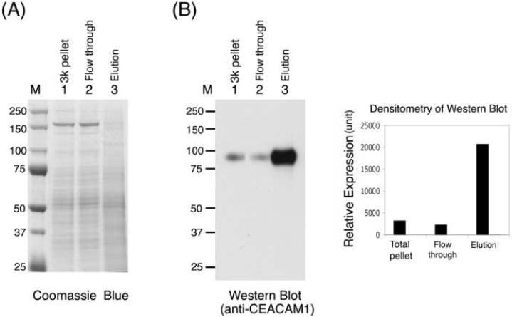
Fig. 3.
Enrichment of plasma membrane from rat liver lysate. The rat liver lysate was prepared as described in Materials and Methods. Equal amounts of protein (4 μg) from the total lysate, flow-through, wash, and elution fractions were loaded on SDS-PAGE gels. The gels were (A) stained with Coomassie blue or (B) transferred to nitrocellulose membranes and immunoblotted with anti-CEACAM1 antibody, and the relative amount of CEACAM1 was determined by densitometric quantification of the signals in the Western blots. M, molecular weight markers. 3k pellet, the pellet fraction from 3,000 xg centrifugation.

