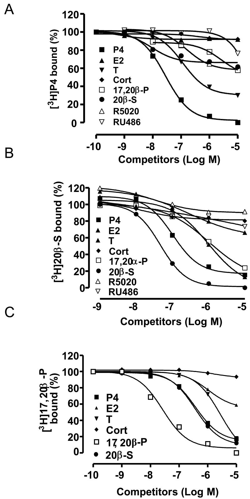
Figure 2.
Competition curves of steroid binding to plasma membranes of MDA-MB-231 (PR-) cells stably transfected with (A) human mPRα, (B) seatrout mPRα, (C) zebrafish mPRα, expressed as a percentage of maximum [3 H]progestin binding (A: [3 H]P4; B:[3 H]20β-S; C: [3 H]17α,20β-P). P4, progesterone; 20β-S, 17,20β,21-trihydroxy-4-pregnen-3-one; 17α,20β-P, 17,20β-dihydroxy-4-pregnen-3-one; RU486, mifepristone; R5020, promegestone; cort, cortisol; E2, estradiol-17β; T, testosterone. (A and B reproduced from Thomas et al., 2007 [122], C reproduced from Hanna et al., 2005 [37] with permission).

